Overview
 Interdigital neuroma (Morton?s Neuroma) of the foot includes common, paroxysmal, neuralgia affecting the web spaces of the toes. It involves entrapment neuropathy (nerve compression) of the common digital nerve below and between the metatarsal heads, typically between the third and the fourth metatarsal heads. The pain is most commonly felt between the third and fourth toes but can also occur in the area between the second and third toes.
Causes
Morton’s neuroma develops for several reasons. The primary reason is wearing narrow toe-box shoes, which compress the metatarsal heads. Certain anatomical factors also make nerve compression more likely with the narrow toe box shoes. In some people fibers, the medial and lateral plantar nerves converge close to the heads of the third and fourth metatarsals. This junction creates a larger nerve structure between the metatarsal heads making it more vulnerable to compression.
Symptoms
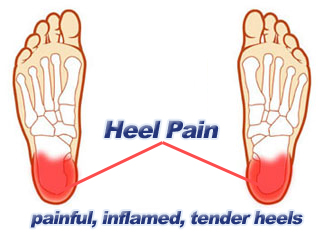
The most common presenting complaints include pain and dysesthesias in the forefoot and corresponding toes adjacent to the neuroma. Pain is described as sharp and burning, and it may be associated with cramping. Numbness often is observed in the toes adjacent to the neuroma and seems to occur along with episodes of pain. Pain typically is intermittent, as episodes often occur for minutes to hours at a time and have long intervals (ie, weeks to months) between a single or small group of multiple attacks. Some patients describe the sensation as “walking on a marble.” Massage of the affected area offers significant relief. Narrow tight high-heeled shoes aggravate the symptoms. Night pain is reported but is rare.
Diagnosis
An MRI scan (magnetic resonance imaging) is used to ensure that the compression is not caused by a tumor in the foot. An MRI also determines the size of the neuroma and whether the syndrome should be treated conservatively or aggressively. If surgery is indicated, the podiatrist can determine how much of the nerve must be resected. This is important, because different surgical techniques can be used, depending on the size and the position of the neuroma. Because MRIs are expensive, some insurance companies are reluctant to pay for them. If the podiatrist believes an MRI is necessary, he or she can persuade the insurance company to pay for it by presenting data to support the recommendation.
Non Surgical Treatment
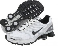
The first step in treating Morton’s Neuroma is to select proper footwear. Footwear with a high and wide toe box (toe area) is ideal for treating and relieving the pain. The next step in treatment is to use an orthotic designed with a metatarsal pad. This pad is located behind the ball-of-the-foot to unload pressure, and relieve the pain caused by the neuroma.
If problem persists, consult your foot doctor.
Surgical Treatment
About one person in four will not require any surgery for Morton’s neuroma and their symptoms can be controlled with footwear modification and steroid/local anaesthetic injections. Of those who choose to have surgery, about three out of four will have good results with relief of their symptoms. Recurrent or persisting (chronic) symptoms can occur after surgery. Sometimes, decompression of the nerve may have been incomplete or the nerve may just remain ‘irritable’. In those who have had cutting out (resection) of the nerve (neurectomy), a recurrent or ‘stump’ neuroma may develop in any nerve tissue that was left behind. This can sometimes be more painful than the original condition.
Prevention
To help reduce your chance of developing Morton’s neuroma avoid wearing tight and/or high-heeled shoes. Maintain or achieve ideal body weight. If you play sports, wear roomy, properly fitting athletic footwear.


 Overview
 Symptoms
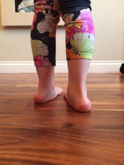 Overview
 Symptoms
 Prevention


 Achilles tendinitis is an overuse injury common in many sports that require lots of running and jumping. Once this condition becomes more chronic adhesions that form along the tissues and the injury becomes more of a tendinosis. Treatment for a tendinosis is much different that for a tendinitis, so it is important to recognize what stage the injury is at in order to treat it appropriately. An acute achilles tendinitis involves inflammation and would be treated with rest, ice, etc. Once the inflammation has decreased, research shows that eccentric exercises are beneficial. Once there is tendinosis, it becomes imperative to break up those adhesions with ART and prescribe appropriate stretches and exercises.

You must be logged in to post a comment.